CRD: component-resolved diagnostics
Molecular testing identifies IgE against individual proteins, not only against whole extracts, and separates primary sensitization from cross-reactivity.
From allergen source to allergen component
Molecular allergens have been described in the scientific literature for more than a decade, but only in recent years have they started to be used more widely in day-to-day allergy practice.
Using allergen components to understand the molecular allergology of patients with a complex or inconclusive sensitization profile on conventional testing has real potential to improve clinical decision-making. Applying component-resolved diagnostics can streamline investigation strategies and improve the diagnosis, the management plan and the advice given to allergic patients.

The sum of sensitizations versus individual proteins
While traditional extract-based serum IgE tests measure the sum of sensitization to all the protein components of a whole allergen, peanut for example, molecular allergology makes it possible to investigate the individual proteins of clinical importance within that same allergen for specific IgE sensitization.
IgE antibody profiles against these molecules vary considerably from one patient to another, and they also differ geographically because of local differences in exposure.
Clinical relevance
Molecular testing with ImmunoCAP™ Component-Resolved Diagnostics allows specific IgE antibodies to be identified against individual allergen molecules, not only against whole allergen extracts. This approach gives a more precise characterisation of the sensitization profile and helps distinguish primary, clinically relevant sensitization from sensitization driven by cross-reactivity.
In allergy practice, CRD testing is particularly useful in:
- polysensitization;
- food allergy with a risk of systemic reactions;
- suspected oral allergy syndrome;
- selecting patients for allergen-specific immunotherapy;
- assessing the risk associated with particular molecular families, such as storage proteins, LTP, PR-10, profilins or polcalcins.
Set in the context of the clinical history and the presenting symptoms, ImmunoCAP™ molecular testing supports a finer allergy diagnosis, better risk stratification and more personalised management.
Allergenic protein families
Introducing molecular allergen testing into routine clinical practice improves allergy diagnosis by working from allergenic proteins and protein families. Protein families are groups with similar function and structure that occur across many allergen sources.
Plant seeds, for example, contain storage proteins such as the vicilins. Lipocalins and serum albumins are examples of protein families found in allergen sources of mammalian origin.

Allergens and allergen components are identified and classified through a partnership between the World Health Organization and the International Union of Immunological Societies. The WHO and IUIS Allergen Nomenclature Sub-Committee maintains a unique, unambiguous and systematic nomenclature for allergenic proteins, based on the Linnaean system.
Specific allergens and cross-reactive allergens
Molecular allergens can be divided into those with a high and those with a low potential to trigger clinical symptoms. They can also be grouped into molecules that are specific to one allergen source and molecules with very similar structures present in different allergen sources. The latter are known as cross-reactive allergens.
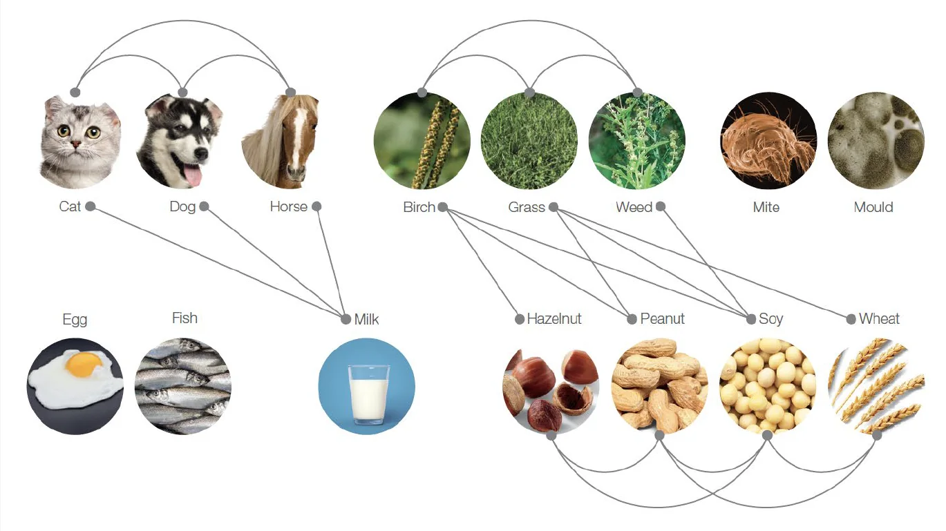
Dog, cat and horse, for instance, contain proteins from the lipocalin family together with serum albumin, which is also found in milk. Birch, grasses and weeds contain profilins, which are also present in legumes such as soy and peanut, as well as in wheat and hazelnut.
Cross-reactive IgE responses can complicate the interpretation of extract-based test results, making it difficult to identify the primary allergen responsible for the symptoms. ImmunoCAP™ Allergen Component tests and the ImmunoCAP™ ISAC multiplex help increase diagnostic clarity.
High and low allergenic potential
Foods are made up of complex matrices of natural components such as proteins, lipids and carbohydrates. Most allergens are proteins, formed of amino acid chains. Within these structures there are regions called epitopes, the recognition sites to which specific IgE antibodies bind.
Molecules with high allergenic potential
Some proteins are more resistant to metabolic processes because of their robust chemical structure, for example the peanut storage proteins (Ara h 1, Ara h 2, Ara h 3, Ara h 6) or ovomucoid from hen’s egg (Gal d 1).
Because they resist digestion better, their epitope structures stay intact for longer, and these proteins can cause systemic symptoms more often than unstable proteins do.
Molecules with low allergenic potential
Molecules such as PR-10 and the profilins, found in nuts, fruit and pollen, are structurally more labile and susceptible to heat, cooking or the action of gastrointestinal enzymes.
They start to break down as early as the oral cavity, causing milder reactions such as oral allergy syndrome. Because the epitope regions are destroyed, these molecules rarely induce systemic symptoms.

In pollen-allergic patients, IgE antibodies initially directed against pollen proteins, Bet v 1 from birch for example, can cross-react with similar proteins in foods. This generates a broad sensitization profile that is regarded as secondary to the pollen sensitization. In clinical practice the phenomenon is known as pollen-food syndrome, and in the context of latex as latex-fruit syndrome.
Cross-reactive allergens also exist in other sources, such as hymenoptera venoms, fish, mites and crustaceans. House dust mites and shrimp, for example, share a cross-reactive protein called tropomyosin.
When sensitization to cross-reactive allergens is identified, the primary sensitizing allergen should be sought in order to understand the mechanism of the patient’s allergy. Testing for both source-specific and cross-reactive allergen components makes it possible, in most cases, to distinguish primary from secondary reactions.